Microscope slides, the cornerstone of biological and non-biological studies, provide invaluable insights into the intricate world of living and nonliving organisms. This article delves into the captivating realm of living vs nonliving microscope slides, exploring their applications, advantages, and disadvantages, while shedding light on their crucial role in scientific research and discovery.
Introduction

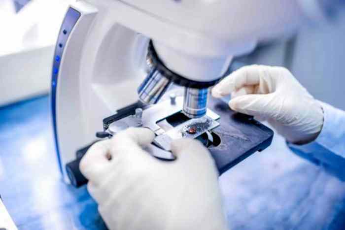
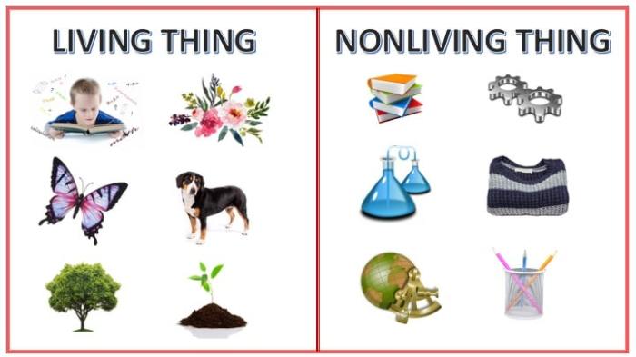
In the realm of microscopy, the distinction between living and nonliving microscope slides plays a pivotal role in biological and non-biological investigations. Living slides, as the name suggests, showcase live specimens, offering a glimpse into the dynamic processes of life.
Nonliving slides, on the other hand, present preserved or inert specimens, providing a more static representation of biological structures or non-biological materials.
The choice between living and nonliving slides hinges on the specific objectives of the study. Living slides are indispensable for observing real-time cellular processes, such as cell division, motility, and interactions with their environment. They are particularly valuable in fields like cell biology, microbiology, and developmental biology.
Nonliving slides, on the other hand, are more suitable for studying the morphology and structure of biological specimens. They are commonly used in histology, anatomy, and pathology to examine tissues, organs, and pathological changes. Additionally, nonliving slides are employed in non-biological disciplines, such as geology, mineralogy, and material science, for analyzing the properties of various materials.
Advantages and Disadvantages
Both living and nonliving microscope slides have their respective advantages and disadvantages.
Advantages of Living Slides:
- Direct observation of cellular processes in real-time
- Study of dynamic interactions between cells and their environment
- Provides insights into cellular physiology and behavior
Disadvantages of Living Slides:
- Limited lifespan of specimens, requiring frequent slide preparation
- Potential for contamination and alteration of specimens during handling
- Specialized equipment and expertise may be necessary for proper preparation and observation
Advantages of Nonliving Slides:
- Preservation of specimens allows for long-term storage and analysis
- Provides a static representation of structures for detailed examination
- Easier to prepare and handle compared to living slides
Disadvantages of Nonliving Slides:
- Loss of cellular function and dynamic processes
- Potential for artifacts and distortions due to preservation techniques
- May not be suitable for studying real-time cellular interactions
Types of Living Microscope Slides

Living microscope slides showcase biological specimens that are still alive and active. These slides offer a unique opportunity to observe cellular processes and organism behavior in real-time. There are various types of living microscope slides, each with its own preparation methods, advantages, and disadvantages.
Wet Mounts
Wet mounts are the simplest type of living microscope slide. They involve placing a drop of liquid containing the specimen directly onto a microscope slide and covering it with a coverslip. Wet mounts are easy to prepare and can be used to observe small, aquatic organisms like paramecia or bacteria.
Advantages:
- Simple and inexpensive to prepare
- Allows for observation of live specimens in their natural environment
Disadvantages:
- Specimens may move or drift, making it difficult to focus
- Limited magnification and resolution
Hanging Drop Slides
Hanging drop slides are used to observe microorganisms that require a liquid environment but also need to be suspended in air. A drop of liquid containing the specimen is placed on a coverslip and inverted over a depression slide. The specimen is suspended in the liquid drop, allowing for observation from multiple angles.
Advantages:
- Provides a stable and enclosed environment for the specimen
- Allows for high-resolution imaging
Disadvantages:
- Can be difficult to prepare and requires specialized equipment
- Specimens may be damaged if the drop dries out
Tissue Cultures
Tissue cultures are slides that contain living cells or tissues that have been grown in a laboratory environment. They are used to study cell growth, differentiation, and interactions. Tissue cultures can be prepared from a variety of sources, including human, animal, and plant tissues.
Advantages:
- Allows for long-term observation of cell behavior
- Can be used to study the effects of different drugs or treatments on cells
Disadvantages:
- Can be expensive and time-consuming to prepare
- Cells may not behave the same way in culture as they do in the body
Types of Nonliving Microscope Slides

Nonliving microscope slides encompass a diverse range of specimens that have undergone specific preparation techniques to preserve their structural integrity and facilitate microscopic examination.
Stained Slides
Stained slides involve treating the specimen with dyes or stains that selectively bind to specific cellular components, enhancing their visibility under a microscope. This technique allows for the visualization of specific structures, such as chromosomes, organelles, and proteins, within the specimen.
Fixed Slides
Fixed slides utilize chemical fixatives to preserve the specimen’s morphology and prevent decomposition. This process involves immersing the specimen in a fixative solution, such as formalin or alcohol, which cross-links proteins and stabilizes cellular structures. Fixed slides provide a more permanent and stable preparation compared to fresh specimens.
Cytology Slides
Cytology slides specifically examine individual cells or cell populations. They are commonly used in diagnostic pathology to detect abnormalities in cell morphology and identify disease processes. Cytology slides are prepared by collecting cells from the body, such as through a Pap smear or biopsy, and then staining and mounting them on a glass slide for microscopic examination.
Applications of Living and Nonliving Microscope Slides: Living Vs Nonliving Microscope Slides

Microscope slides play a crucial role in various scientific disciplines, offering insights into the microscopic world. Both living and nonliving microscope slides serve distinct purposes and have specific advantages and disadvantages.
Applications of Living Microscope Slides
Living microscope slides provide a unique opportunity to observe living organisms in their natural state, allowing researchers to study their behavior, physiology, and interactions in real-time. These slides are commonly used in:
- Biology:Examining cellular processes, such as mitosis, meiosis, and cell division.
- Medicine:Diagnosing diseases by analyzing blood, urine, or tissue samples.
- Environmental science:Studying microorganisms in water, soil, and other ecosystems.
Applications of Nonliving Microscope Slides
Nonliving microscope slides are used to examine non-living specimens, providing detailed structural and morphological information. They are widely employed in:
- Histology:Studying the microscopic structure of tissues.
- Pathology:Diagnosing diseases by examining tissue biopsies.
- Forensics:Analyzing trace evidence, such as fibers, hair, and fingerprints.
Comparison of Living vs Nonliving Microscope Slides
The choice between living and nonliving microscope slides depends on the specific application and the desired information. Living slides offer real-time observations of biological processes, while nonliving slides provide detailed structural information and allow for long-term storage.
Advantages of Living Microscope Slides:
- Direct observation of living organisms
- Study of dynamic processes
- Analysis of interactions between organisms
Advantages of Nonliving Microscope Slides:
- Detailed structural information
- Long-term storage and preservation
- Easier preparation and handling
Conclusion

In conclusion, living and nonliving microscope slides are essential tools for studying the microscopic world. Living microscope slides provide a unique opportunity to observe living organisms in their natural state, while nonliving microscope slides allow for the detailed examination of preserved specimens.
The future of microscopy holds exciting possibilities, with advancements in technology enabling the development of new techniques and applications. Further research and exploration are needed to fully unlock the potential of microscopy in various fields of science and medicine.
Future Directions in Microscopy, Living vs nonliving microscope slides
The future of microscopy is bright, with several promising directions for research and development. These include:
- Super-resolution microscopy:Techniques like stimulated emission depletion (STED) and photoactivated localization microscopy (PALM) are pushing the limits of resolution, allowing for the visualization of structures at the nanoscale.
- Live-cell imaging:Advancements in fluorescence microscopy and optogenetics are enabling the real-time visualization of cellular processes in living organisms.
- Correlative microscopy:Combining different microscopy techniques, such as electron microscopy and light microscopy, provides a comprehensive view of biological structures at multiple scales.
- Artificial intelligence (AI):AI algorithms are being applied to microscopy data to automate image analysis, identify patterns, and make predictions.
Suggestions for Further Research and Exploration
To further advance the field of microscopy, several areas warrant further research and exploration:
- Development of new imaging modalities:Exploring novel approaches to microscopy, such as X-ray microscopy and atomic force microscopy, can provide complementary information and insights.
- Optimization of sample preparation techniques:Improving methods for preparing and preserving biological specimens can enhance image quality and reduce artifacts.
- Integration of microscopy with other technologies:Combining microscopy with techniques like genomics, proteomics, and metabolomics can provide a more holistic understanding of biological systems.
- Educational outreach:Promoting microscopy education and training can inspire future generations of scientists and researchers.
Query Resolution
What is the primary difference between living and nonliving microscope slides?
Living microscope slides contain living organisms or tissues, while nonliving slides showcase preserved or stained specimens.
Which type of microscope slide is more suitable for studying cellular processes?
Living microscope slides are ideal for observing dynamic cellular processes, as they allow researchers to witness real-time biological activity.
What are the advantages of using nonliving microscope slides?
Nonliving slides offer greater longevity, reduced risk of contamination, and the ability to capture permanent images for documentation and analysis.
